
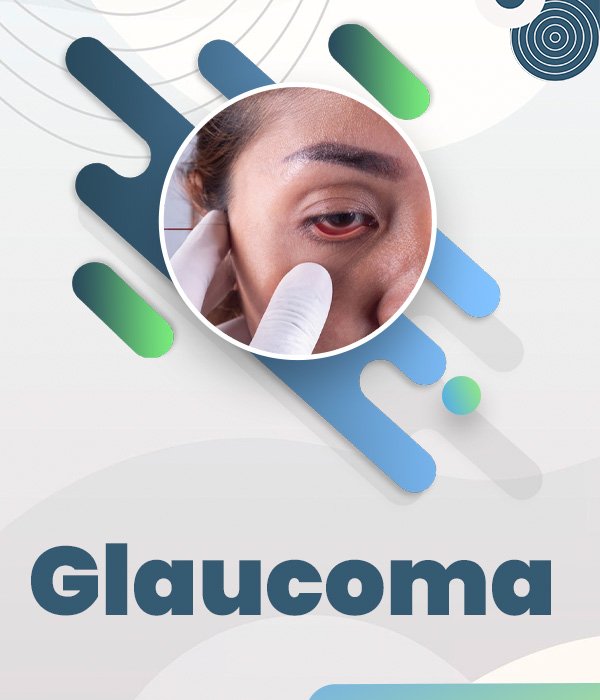
What is Glaucoma?
A layer of the retina that is typically caused by an elevation in the intraocular pressure. It can go undetected until the extreme visual loss has taken place. People at risk for glaucoma include those with a family history of glaucoma, diabetics, those with hypertension, individuals of African-American decent, and those of increasing age. In general, most glaucoma is painless and asymptomatic. The visual loss that initially occurs is in the periphery of the visual field, so most people do not even notice the visual loss.
As glaucoma progresses, and if left
untreated, it can affect the central vision in an irreversible fashion. Glaucoma is diagnosed on a routine screening eye examination. The initial treatment for glaucoma is the use of eye drops in order to reduce intraocular pressure. If eye drops fail, then laser or even surgical repair is necessary. That’s why it is crucial for an individual to undergo an annual eye examination, especially if there is a risk factor for such a blinding disease.
Symptoms of Glaucoma
Symptoms are:
(a) Frequent change of glasses (none of which is satisfactory)
(b) Difficulty in dark adaptation.
(c) Bumping into things in unfamiliar places.
(d) Blurred or foggy vision.
(e) Coloured halos (rainbow-colored rings) around lights.
More detailed information about Glaucoma

Glaucoma - “The silent thief of Sight”
Just imagine a class of 100 people graduating from class 10 at age 15. If we check their eye pressure, maybe 2 or 3 would have elevated eye pressure. If the same group of people met after 2 decades and we checked their pressure again at least 5 to 7 would have elevated pressure. If they were all alive at the age of 75 i.e. 4 decades later, at least 20 of them would have developed glaucoma. Recent studies show that Glaucoma develops with increasing age to the tune of 2% per decade of life.
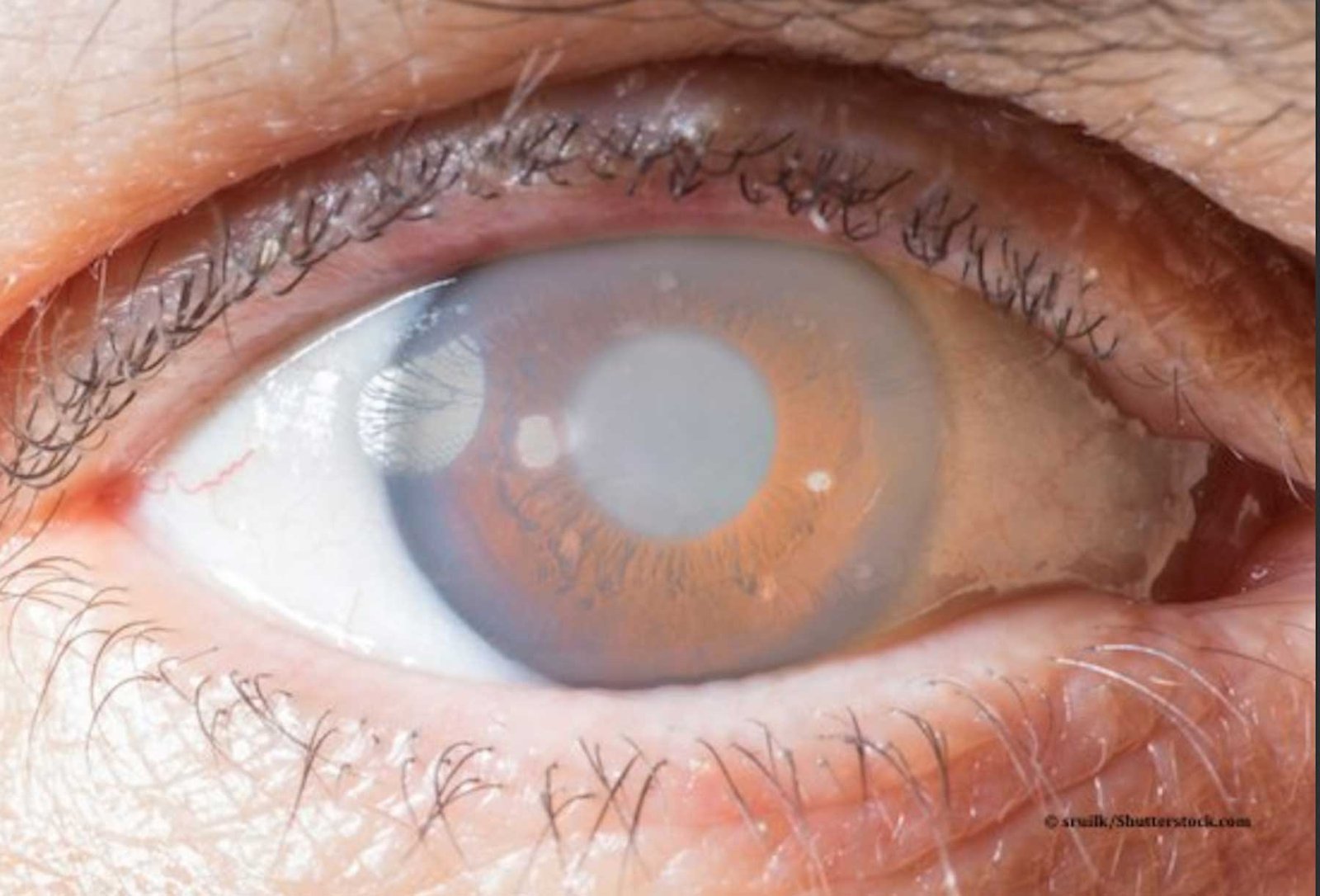
Glaucoma is not a new ailment. It was described hundreds of years ago by Greek, Arab and Indian surgeons who told of a “tightness” and “whitening” of the eye, of a “black water”, that filled inside the eye robbing the patient of sight. To this day across India, people refer colloquially to this as “Kala Motia” or in Gujarati as “Jhamar.”
The human eye has a part called the ciliary body that produces a fluid called “Aqueous”. This aqueous has to flow out of the eye through a mesh similar to a tea strainer. With age, injury, diabetes, swelling, cataract etc the mesh gets blocked leading to rising in pressure in the eye. This rise in pressure presses on the optic nerve inside the eye.
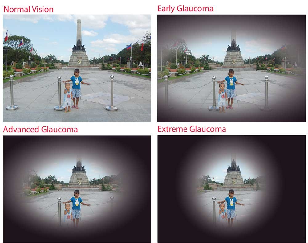
The peripheral vision (side vision) decreases. However, there is usually no pain and central vision is preserved to the end. Since central vision and reading vision are preserved until the disease is very severe, it usually slips the patient’s attention and is detected accidentally by the ophthalmologist when the person visits them for some other eye complaint.
Glaucoma there for must consist of 3 basic findings
1. Increased pressure in the eye (normal is usually taken as less than 21 millimeters of mercury).
2. Optic nerve changes which are seen by your eye doctor by looking inside the eye with an instrument.
3. Loss of “field” or side vision.
The pressure can be checked by a machine called a tonometer. Modern set-ups have a computerized device that blows a puff of air at the eye to measure its pressure. Field testing is done on a large unit called an “Automated Perimeter”. This machine shows spots of light at various places on a screen. Inability to see these spots are noted by the machine as field loss and a map is plotted by the computer.
In the very late stage of the disease, the patient feels that he or she is looking through a tube as the side vision is gone. This is called tubular vision.
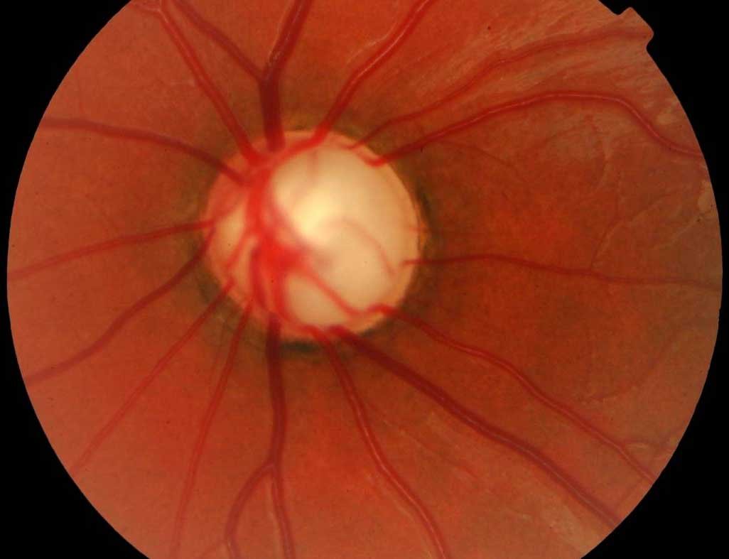
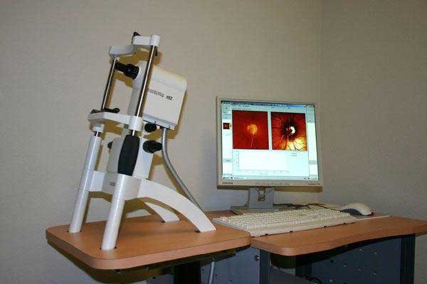
The optic nerve changes today can be measured by a machine called an HRT-2 from Germany, which measures the changes and gives a report. This is performed every 4-6 months to know if the nerve changes are progressing or not. In the olden days, the eye doctor would look in and draw a diagram, however today this machine plots for us 69,000points on the nerve which is as small as 3 mm in diameter giving us a much more accurate assessment.
However modern technology or not it cannot replace the doctor’s experience. Remember the old saying, that, “In a car accident the loosest nut is the one behind the wheel”. HRT-2 has been proven to detect glaucoma up to 5 years before the field test and is our new warrior in the fight against Glaucoma.
In some cases, glaucoma can be there without increased pressure, called Normal Tension Glaucoma (NTG). It is now known that more than 40% of all glaucoma patients are NTG patients. These normal tension glaucoma patients’ eyes cannot stand even normal pressure and the pressure needs to be reduced by 30% lower than it is. For example, if a patient has 18 pressure and is diagnosed by the eye doctor with normal tension glaucoma the pressure will have to be reduced to 12 to control the progress of the disease.
Glaucoma is not just a disease of old age. Newborn babies can have glaucoma as a congenital problem. When pressure rises in a baby’s eye the eye swells up and looks huge like an ox or buffalo eye. This appearance is called “Bupthalmos”. If the child is treated with the correct surgery before the age of 3 the eye usually returns to its normal size. If as is usually the case in rural areas the child does not receive the correct surgical treatment the eye becomes very large and vision is greatly reduced leading to blindness!
Glaucoma and eye pressure rising can also be due to many other causes. Bleeding inside the eye can lead to pressure rising. Diabetes can cause a pressure rise. A mature cataract can cause a pressure rise. Any blow to the eye can lead to a rise in pressure even 6 months after the injury. Pressure can go up as a complication or a sequel to cataract surgery and was more common in the past when the eye was cut open and sutured.
A famous case of pressure rising in the eye is the use of corticosteroid medicines. Patients who use steroid-containing skin creams, eye drops and tablets or injections are at risk of rising eye pressure. All these glaucomas which are due to other causes are called “Secondary Glaucoma”.
Finally, glaucoma can sometimes be an emergency when due to the mesh getting totally blocked the pressure suddenly goes to 50 and above. Typically the patient will complain of pain watering and redness and poor vision. This type of acute glaucoma “Attack” is to be aggressively treated with IV injection, laser and eye drop otherwise it will lead to blindness.
Diagnosis... State of the Art!
The Heidelberg Retina Tomograph or HRT is the finest technique in the early detection, evaluation and checking progression of Glaucoma. It is a confocal scanning laser imaging device that obtains three-dimensional images of the optic nerve head and retina in the posterior segment, checking the height changes or differences in the optic nerve head. There is now good evidence that the HRT can diagnose glaucoma 5 years before changes become evident by any other technique. The HRT is now the gold standard for imaging the optic nerve. The Mehta International Eye Institute uses the Heidelberg Retina Tomograph in its continuous effort to provide quality care to its patients. The test is usually repeated thrice a year and helps the physician assess if changes are occurring in the nerve fiber layer or the optic nerve head. These changes almost always occur prior to any visual symptoms in the patient.
The Humphrey Computerised Field analyzer
An instrument that evaluates what you can see with your “fields” from the periphery of your eye while keeping the eye fixated in the middle. It evaluates the extent of the loss. In glaucoma, the fileds progressively decrease till only a central island is left, which if untreated, extinguishes leading to blindness. The Humphrey Analyzer is the Gold standard all over the world. Fields are normally done twice a year.

Intraocular pressure evaluation
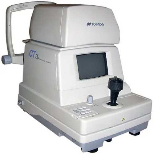
Non-Contact Pneumotonometer: Acts as an analyzing tool and is excellent as a survey instrument. Reasonably accurate when kept properly gradted. The Topcon is perhaps the most sensitive of this genere.
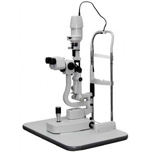
Goldman Applanation Slit Lamp Biomicroscope mounted Tonometer
Exceptionally accurate but requires steady fixation. Can identify pressure accurately even in post-operative and highly myopic eyes.
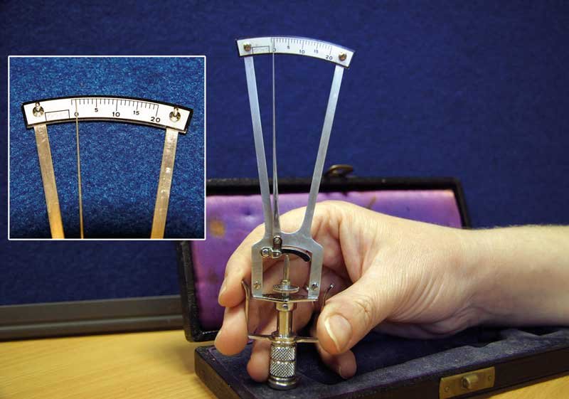
Schiotz Tonometry
When the surface is grossly irregular or as a verification device the Schiotz tonometry is an exceptional tool.
Treatment for Glaucoma
Glaucoma can be treated medically or surgically. Early glaucoma is usually easily treated with just eye drops. These eye drops will have 2 actions:
1. Reduce the amount of fluid produced in the eye and or
2. Speed up the exit of fluid from the eye by action on the meshwork and other pathways.
Most glaucoma eye drops cause a little bit of irritation and dryness of the eye along with redness and must be used under the doctor’s orders only. Some eye drops like Timolol can aggravate a patient’s asthma and some like Prostaglanding cause the skin around the eye to darken and eyelashes in that eye to grow. Do speak with your eye doctor about the side effects of glaucoma eye drops. Rarely when eye pressure does NOT come under control Diamox tablets prescribed. A common side effect is tingling and numbness in the hands and feet.
Laser: In case there is an Attack of glaucoma it can be controlled by application of YAG: Laser SLT is a new laser that can treat chronic simple glaucoma as well.
Surgery: Since the seventies the most common surgery for glaucoma is Trabeculectomy. In this a flap is made in the white of the eye and under that, a hole is cut into the eye from which the fluid flows out. Problems with this were bleeding, very low pressure and poor vision after surgery. Such problems led to patients fearing glaucoma surgery and led to many patients losing their sight rather than opting for this surgery.
In the late nineties, a new procedure came out in Switzerland and western Europe called Non-Penetrating Deep Sclerectomy. Here the eyeis NOT OPENED and only the sclera i.e. the white of the eye is thinned until the fluid canal called Canal of Schlemm is opened. The
advantage with this is that the pressure is maintained low and there is no chance of bleeding, poor vision, too low pressure etc. This procedure now is the glaucoma surgery of choice in the west. In India, it still has to catch on. The author began performing this surgery back in 2001 and has reported excellent results with it.


